
Welcome to Amanda's NICU Education


Hi! My name is Amanda. I'm a NICU nurse, Clinical Nurse Specialist, NICU Educator... basically your NICU BFF. If you want to talk NICU, I'm here for you! I love everything about NICU nursing and I'm eager to learn and share my knowledge with all my NICU friends.
I have been a NICU nurse since 2009 I am currently a Clinical Nurse Specialist in a Level IV NICU in Los Angeles.
I am passionate about educating the next generation of NICU nurses. I share my knowledge through platforms such as Instagram and Facebook and am excited to have you here on my website!
Click on the button below to sign up for my newsletter filled with NICU education and tips for all experience levels.

Not very many people love taking tests but as a self-acclaimed "forever student" who has taken (and passed) five different certification exams I am no longer afraid of tests! "Way to brag", you might be thinking but I want to help YOU pass your certification exam too!
Introducing Amanda's RNC-NIC Success digital course - your ultimate study companion!
Gain unlimited, on-demand access for life, ensuring you're primed to ace your certification exam.
I'm here to help you succeed and I can't wait for you to share with me that you PASSED the RNC-NIC EXAM!!!


Neonatal Arrythmias
Arrhythmias in the NICU: What Nurses Need to Know to PASS the RNC-NIC
Cardiac rhythm disturbances are not the most common problem in the NICU, but when they occur they can become critical quickly. For NICU nurses, recognizing abnormal rhythms early and understanding the underlying physiology can make a significant difference in outcomes.
Arrhythmias in neonates range from benign rhythm variations to life-threatening tachyarrhythmias or conduction abnormalities. Because newborn physiology is unique, the presentation and management of arrhythmias can differ significantly from older pediatric or adult patients.
In this blog, we'll review:
How arrhythmias present in the NICU
Common causes of neonatal arrhythmias
How they are diagnosed and managed
Potential complications and outcomes
Presentation and Assessment of Arrhythmias in the NICU
The challenge with neonatal arrhythmias is that they may present subtly. Many infants initially show nonspecific signs of cardiovascular compromise rather than obvious rhythm disturbances.
Common clinical signs
NICU nurses may notice:
Tachycardia or bradycardia
Feeding intolerance
Poor perfusion
Pallor or cyanosis
Irritability or lethargy
Apnea or desaturation events
Signs of heart failure
In severe cases, infants may develop:
Hypotension
Decreased urine output
Metabolic acidosis
Shock
Rhythm assessment
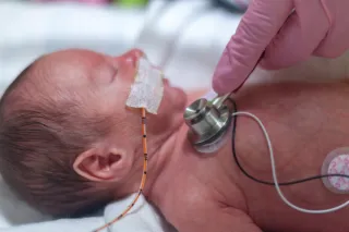
Assessment usually begins with bedside monitoring.
Important steps include:
Continuous ECG monitoring
Assessing heart rate trends
Evaluating perfusion and blood pressure
Reviewing telemetry rhythm strips
Obtaining a 12-lead ECG
Monitoring oxygen saturation and perfusion
Medical providers may also order:
Electrolytes
Blood gas
Echocardiography
Holter monitoring
Careful rhythm interpretation is essential because neonatal heart rates normally range between 120–160 beats per minute, which can make tachyarrhythmias harder to recognize initially.
Causes of Arrhythmias in Neonates
Arrhythmias can arise from abnormalities in the heart’s electrical system or from systemic conditions affecting cardiac conduction.
Common causes include
Structural heart disease
Congenital heart defects can disrupt normal conduction pathways and predispose infants to arrhythmias.
Examples include:
Ebstein anomaly
Atrioventricular septal defects
Congenital corrected transposition
Electrolyte abnormalities
Electrolyte disturbances can significantly alter cardiac conduction.
Examples include:
Hypokalemia
Hyperkalemia
Hypocalcemia
Hypomagnesemia
Hypoxia and ischemia
Perinatal asphyxia or severe respiratory failure can impair myocardial function and trigger rhythm disturbances.
Medication exposure
Certain medications may provoke arrhythmias, including:
Catecholamines
Digoxin
Antiarrhythmics
Some antibiotics
Macrolides (e.g. erythromycin) and Fluoroquinolones (e.g. levofloxacin, moxifloxacin) can cause QT prolongation
Genetic or conduction disorders
Inherited conditions may also affect cardiac conduction, such as:
Long QT syndrome
Congenital heart block
Wolff-Parkinson-White syndrome
Maternal autoimmune disease (especially lupus with anti-Ro/SSA antibodies) is a well-known cause of congenital heart block.
Common Types of NICU Arrhythmias
Sinus tachycardia
The most common tachycardia in neonates.
Causes typically include:
Fever
Pain
Hypovolemia
Infection
Hypoxia
Treatment focuses on correcting the underlying cause rather than treating the rhythm itself.
Supraventricular tachycardia (SVT)
SVT is the most common pathologic tachyarrhythmia in neonates.
Typical heart rates are 220–300 bpm with a narrow QRS and a regular R-R interval.
this helps distinguish SVT from ventricular tachycardia, which produces wide QRS complexes.
Signs may include:
Poor feeding
Irritability
Tachypnea
Pallor
Heart failure if sustained
Atrial flutter
Less common but important to recognize.
Atrial rates may exceed 300 beats per minute, often with a 2:1 conduction pattern. The classic ECG finding in atrial flutter is the “sawtooth” pattern created by continuous atrial depolarization.
Premature atrial or ventricular contractions
These are usually benign and transient in neonates.
They often resolve without treatment but should still be monitored.
Congenital heart block
Congenital heart block can occur when maternal autoantibodies cross the placenta and damage the fetal cardiac conduction system, particularly the AV node.
The most important antibodies to know are:
Anti-Ro (SSA) antibodies
These are the most commonly associated antibodies with congenital heart block.
They are commonly found in mothers with:
Systemic lupus erythematosus (SLE)
Sjögren syndrome
However, some mothers are asymptomatic carriers and may not know they have these antibodies.
Congenital heart block is part of a broader condition called neonatal lupus syndrome, which can include:
Congenital heart block
Transient rash (usually resolves by 6-8 months)
Sometime described as "Raccoon" or "Owl" Eyes as periorbital edema is a classic finding
The rash can also present on the face, scalp, trunk, and extremities
Liver dysfunction
Pancytopenia:
Thrombocytopenia (most common)
Anemia
Neutropenia
The heart block is the most serious and often permanent complication.
Management of Neonatal Arrhythmias
Management depends on the type of arrhythmia and the infant’s hemodynamic stability.
Initial stabilization
The first priority is always assessment of airway, breathing, and circulation.
NICU nurses should anticipate:
Oxygen support
Establishing IV access
Continuous monitoring
Blood gas and electrolyte evaluation
Vagal maneuvers
For stable infants with SVT, providers may attempt vagal stimulation such as:
Ice to the face (diving reflex)
This may temporarily slow conduction through the AV node.
Pharmacologic treatment
Medications frequently used include:
Adenosine
First-line medication for SVT.
It temporarily blocks AV node conduction and may terminate the arrhythmia. Adenosine must be given by rapid IV push, followed by a saline flush as close to the heart as possible. It has a very short half life.
Antiarrhythmics
Examples include:
Propranolol
Amiodarone
Procainamide
These may be used for recurrent or refractory arrhythmias.
Electrical cardioversion
If the infant is unstable, synchronized cardioversion may be required.
This is typically performed in urgent situations of hemodynamic instability.
Long-term management
Some infants may require:
Chronic antiarrhythmic therapy (e.g. propranolol, digoxin, amiodarone, flecainide, Sotalol)
Cardiology follow-up
Pacemaker placement for severe conduction abnormalities
Complications of Neonatal Arrhythmias
Untreated arrhythmias can lead to serious complications.
Potential complications include:
Congestive heart failure
Poor systemic perfusion
Shock
Myocardial dysfunction
Hydrops fetalis (in fetal arrhythmias)
Sustained tachyarrhythmias can also lead to tachycardia-induced cardiomyopathy, where prolonged rapid heart rates weaken the heart muscle.
Outcomes and Prognosis
The good news is that many neonatal arrhythmias resolve with treatment.
For example:
SVT often resolves during infancy
Premature beats typically disappear without intervention
Some conduction disorders improve over time
However, infants with:
Structural heart disease
Genetic arrhythmia syndromes
Severe conduction defects
may require lifelong cardiology care.
Early recognition and treatment significantly improve outcomes.
Key Takeaways for NICU Nurses
Arrhythmias in neonates can present subtly but progress rapidly. NICU nurses play a critical role in identifying abnormal rhythms and initiating timely interventions.
Important points to remember:
Always evaluate perfusion, not just heart rate
Review telemetry strips carefully
Consider reversible causes such as electrolytes or hypoxia
Recognize SVT as the most common neonatal tachyarrhythmia
Escalate concerns early when rhythms change
Want to Feel Confident Recognizing NICU Emergencies?
Understanding cardiac physiology, arrhythmias, and neonatal shock is essential for both bedside care and the RNC-NIC or CCRN-N certification exam.
In my Neonatal Certification Review Course, I break down complex topics like:
Neonatal cardiac physiology
Arrhythmias and congenital heart disease
Vasoactive medications
Neonatal shock and resuscitation
so they actually make sense and can be applied at the bedside.
👉 Explore the course here: https://amandasnicuconsulting.com/rnc-course
Hundreds of NICU nurses have used this course to pass their certification—and many say it’s helped them feel more confident caring for critically ill babies.


December 2023 Certification Review Webinar
NICU Certification Review


Ready to kickstart your journey to becoming a certified NICU nurse?
Look no further!
Grab my FREE E-Book packed with essential study and test-taking strategies for the RNC-NIC.
In the E-Book I give you the resources you need including the link to access the candidate guide, several types of books to study from, some of my favorite strategies, an outline of the content you should review, and a blank calendar for you to make your study plan!
Frequently Asked Questions About the RNC-NIC exam

What is the RNC-NIC?
The RNC-NIC is a competency-based exam that tests the specialty knowledge of nurses in the United States & Canada who care for critically ill newborns and their families.
The RNC-NICU is a nationally recognized certification that recognizes the registered nurse for their specialty knowledge and skill.

Who can take the RNC-NIC exam?
Nurses can take this exam after a minimum of two years experience in the NICU caring for critically ill newborns and their families.

Which books should I use?
I'm glad you asked! There are many excellent books to help you prepare for the RNC-NIC, I gathered ande describe each of them for you in my FREE e-book.
Is there a course to help me study?
Yes! Many hospitals host their own certification course and there are a few online courses. See my RNC-NIC test taking tips E Book for more information
What happens if I don't pass the exam?
If you don't pass the exam on your first try you can try again after 90 days. You will have to reapply after 90 days and pay a retest fee. There is no limit to the number of times you can take the exam (however a candidate can only sit for the exam twice per year).
Can I make more money if I take the RNC-NIC exam and get certified?
Yes! Many hospitals provide a raise or a bonus for nurses with specialty certifications. Hospitals also typically hire at a higher base salary when nurses have a certification.

Find me @amandasnicued on these channels or Email me
hey nurses don't miss out
© Copyright 2024. AmandasNICUEd. All rights reserved. | Terms & Conditions | Privacy Policy Contact: [email protected]